The Laser Photodisruptor is one of the key ophthalmological devices which today has become a necessary medical laser tool for ophthalmology clinics. Using this device, two common eye surgery, capsulotomy and iridotomy and more recently laser vitreolysis are performed as a non-invasive treatment of secondary cataract, angle closure glaucoma and floaters respectively. As the laser-tissue interaction mechanism which caused by said device is photodisruption, it is called laser phodisruptor. The demand for this system is high enough that in spite of the usage of foreign products in the country, it is yet economically sensible to produce in the nation. In addition, fabrication of such devices considered as a key step which would lead to indigenization of related technologies. Laser photodisruptor is consisted of two main parts: Horizontal slit lamp and laser delivery pack. Slit lamp is the base for laser delivery pack and provides illumination light and microscope for inspecting eye interiorly while doing surgery. In fact laser delivery pack would couple to slit lamp to enhance it with two laser sources, one for aiming and the other for treatment. The aiming laser is a red He-Ne and the treatment laser is Nd:YAG with wave length of 1064nm.
|
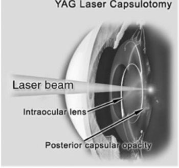
| Laser Posterior capsulotomy |
In one of the works in LO Lab, the design and fabrication process of the said device was performed. In fact design, optical simulation, optical element selection, optical layout setup fabrication and testing and measurements of the laser delivery pack as well as providing suitable slit lamp in order to fabricate such device were carried out. By designing and optical simulating, the optimized layout of the laser delivery pack was determined and the proper elements were selected, purchased and mounted on the system. The design process had two phases, one which roughly gave some information for general layout of the laser system and the other which precisely determined the eligible elements and even their orientations in the layout in order to cancel optical aberrations. Additionally an optical simulation with Zemax software as a complement confirmed the design and also helped with extra information about the best layout. In order to evaluate different layouts, parameters of optical path difference, aberration and spot size were the ones which examined.
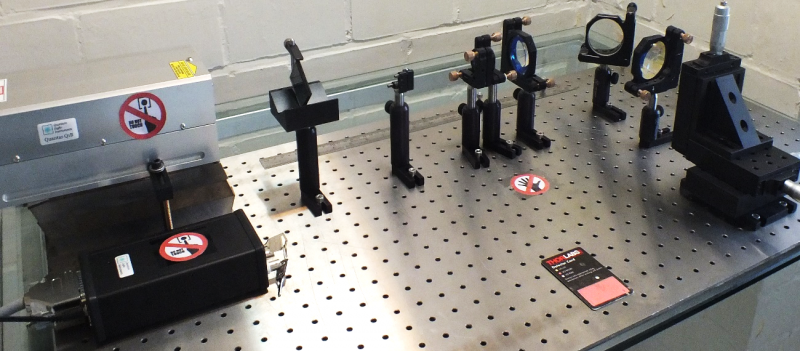
According to simulations results, laser system elements were selected; laser source was chosen so that it met the needs for photodisruption of target tissues. Other elements were selected while their substrate, coating, geometry and laser-induced damage threshold were diagnostic criteria. Finally critical output parameters in accordance with the standards were tested and measured. Beam diameter, divergence angle, spatial profile, repetitions rate, pulse duration and pulse energy were measured with corresponding measuring instruments. Measurements and tests of adjusted layout for laser delivery pack confirmed favorable performance and safe application of the device for the purpose of expected eye surgeries. Plasma formation was observed in target at focal point of the objective lens of the laser system.
|
| Setup for laser beam evaluation |
This study has been done by Mahdi Dehghani.